Home

străpunge În aer liber copleșitor tunica muscularis internetul Strans Punctualitate

Digestive
Digestive

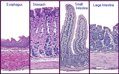
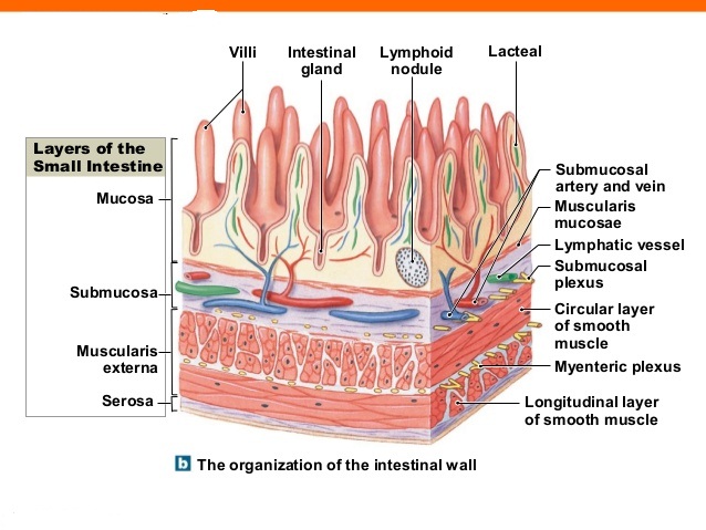
Microanatomy of the Digestive Tube

Fat deposition in the tunica muscularis and decrease of interstitial cells  of Cajal and nNOS-positive neuronal cells in the aged rat colon | American  Journal of Physiology-Gastrointestinal and Liver Physiology
Fat deposition in the tunica muscularis and decrease of interstitial cells of Cajal and nNOS-positive neuronal cells in the aged rat colon | American Journal of Physiology-Gastrointestinal and Liver Physiology
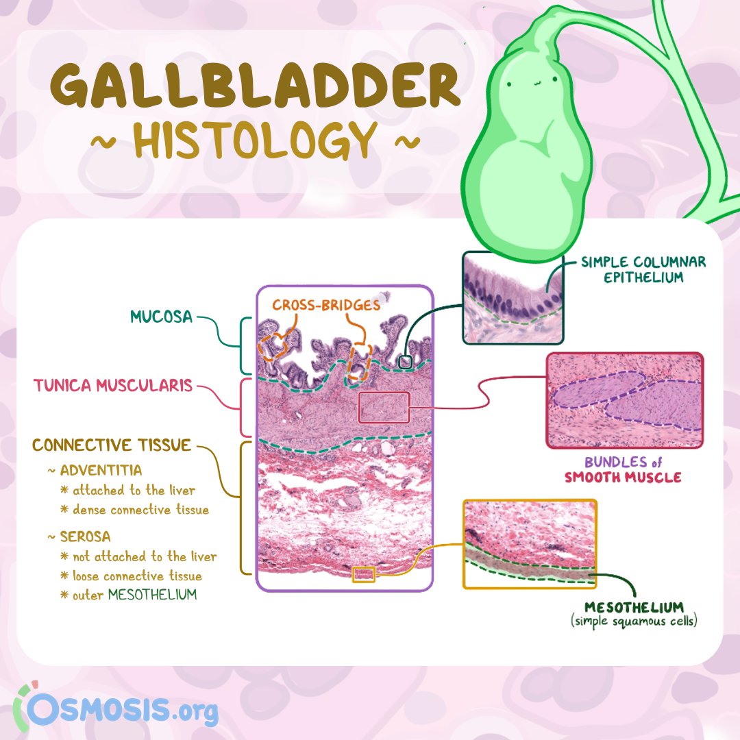
Osmosis from Elsevier on X: "The gallbladder is a muscular pear-shaped  organ that stores, concentrates, and then releases bile into the duodenum.  It has three main layers: the mucosa, tunica muscularis, and
Osmosis from Elsevier on X: "The gallbladder is a muscular pear-shaped organ that stores, concentrates, and then releases bile into the duodenum. It has three main layers: the mucosa, tunica muscularis, and
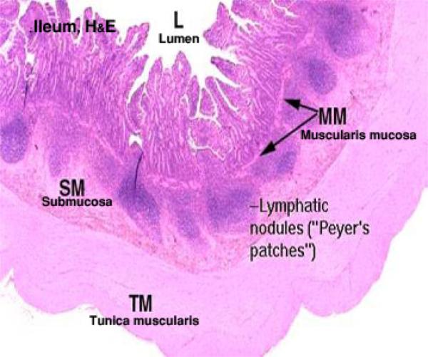
Histology Ileum Diagram | Quizlet

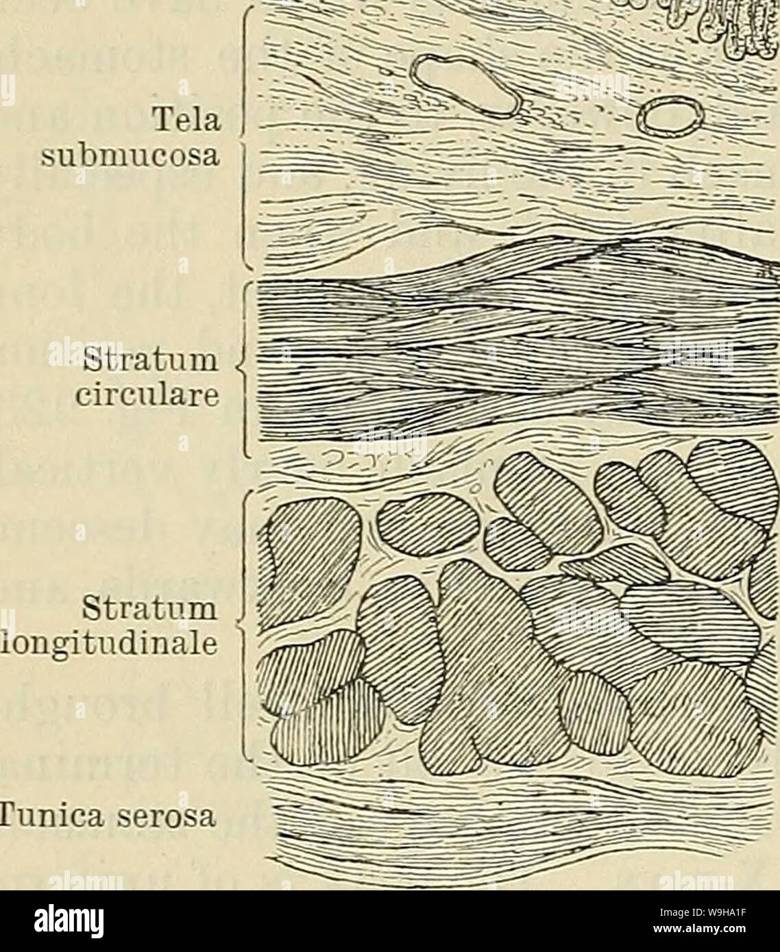
Cunningham's Text-book of anatomy. Anatomy. 1174 THE DIGESTIVE SYSTEM.  il^r^ffli^ Tunica mucosa Tela submucosa Structure of the Stomach. The  stomach wall is composed of four coats—namely, from without inwards: (1)  Tunica
Cunningham's Text-book of anatomy. Anatomy. 1174 THE DIGESTIVE SYSTEM. il^r^ffli^ Tunica mucosa Tela submucosa Structure of the Stomach. The stomach wall is composed of four coats—namely, from without inwards: (1) Tunica

Digestive
Digestive

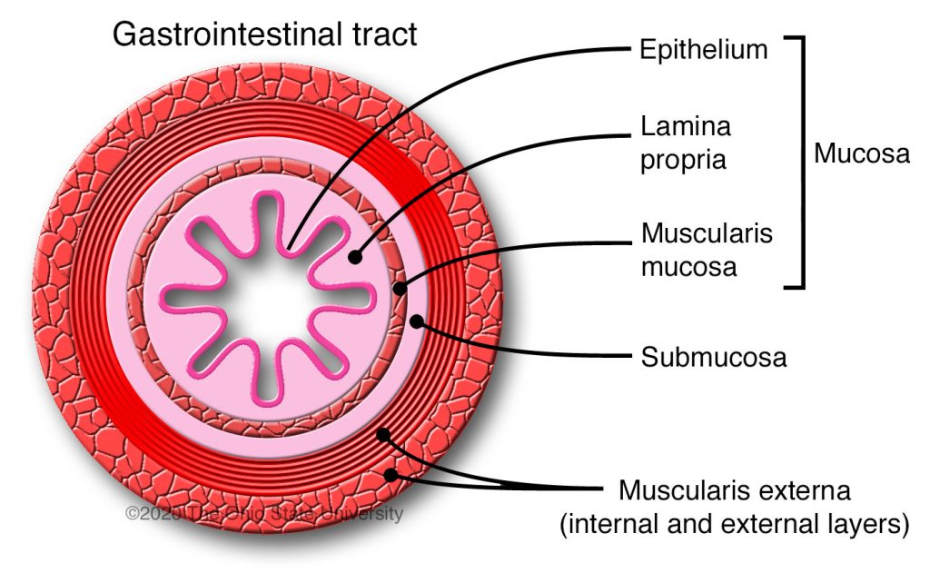
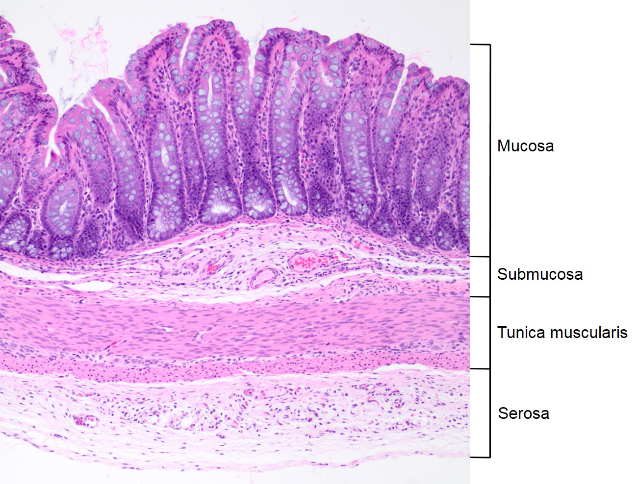
General histologic anatomy of the tubular digestive tract – Veterinary  Histology
General histologic anatomy of the tubular digestive tract – Veterinary Histology

MICROSCOPIC HISTOLOGY IMAGES - GUT

Digestive
Digestive

Animals | Free Full-Text | Morpho-Histological Studies of the  Gastrointestinal Tract of the Orange-Rumped Agouti (Dasyprocta leporina  Linnaeus, 1758), with Special Reference to Morphometry and Histometry
Animals | Free Full-Text | Morpho-Histological Studies of the Gastrointestinal Tract of the Orange-Rumped Agouti (Dasyprocta leporina Linnaeus, 1758), with Special Reference to Morphometry and Histometry

Stomach Diagram | Quizlet

Muscular layer - Wikipedia

From the lumen outward, what are the layers of the gastrointestinal tract?  | Socratic
From the lumen outward, what are the layers of the gastrointestinal tract? | Socratic

Tunica Mucosa - Lamina Propria - Lamina Muscularis - Tunica Submucosa -  Tunica Serosa | PDF
Tunica Mucosa - Lamina Propria - Lamina Muscularis - Tunica Submucosa - Tunica Serosa | PDF

TUBULAR & VISCERAL ORGANS Flashcards | Quizlet

Muscular layer of large intestine - e-Anatomy - IMAIOS

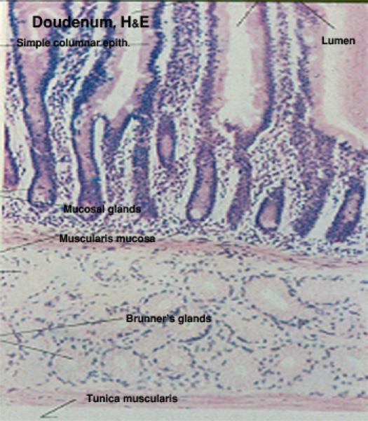
Section of duodenum (European Roller) shows : muscularis mucosa (1),... |  Download Scientific Diagram
Section of duodenum (European Roller) shows : muscularis mucosa (1),... | Download Scientific Diagram

General histologic anatomy of the tubular digestive tract – Veterinary  Histology
General histologic anatomy of the tubular digestive tract – Veterinary Histology

Daily Anatomy - Cross section through the jejunum The jejunum is the middle  section of the small intestine. The jejunum lies between the duodenum and  the ileum. The lining of the jejunum
Daily Anatomy - Cross section through the jejunum The jejunum is the middle section of the small intestine. The jejunum lies between the duodenum and the ileum. The lining of the jejunum
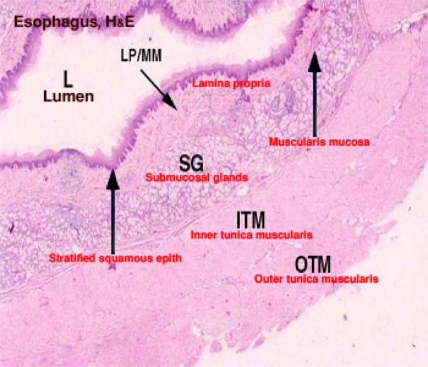
PPT - The General Plan PowerPoint Presentation, free download - ID:1711937

Muscular layer of ureter - e-Anatomy - IMAIOS

Cunningham's Text-book of anatomy. Anatomy. Epithelium Papilla J Conn,  tissue . if Lj I Lamina. §^0ti^^- Tunica mucosa Muscularis mucosas Loose  conn, tissue . Glandula cesophagea Circular â muscle fibres Tela
Cunningham's Text-book of anatomy. Anatomy. Epithelium Papilla J Conn, tissue . if Lj I Lamina. §^0ti^^- Tunica mucosa Muscularis mucosas Loose conn, tissue . Glandula cesophagea Circular â muscle fibres Tela

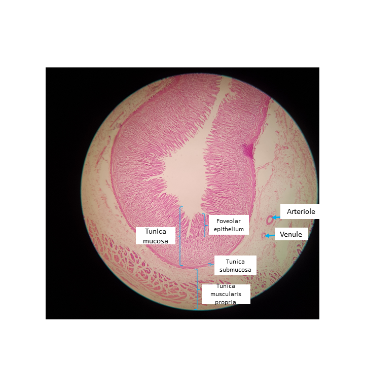
Solved Hi! I'm trying to identify the parts in the fundus | Chegg.com